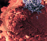
Diagnostic Aspects Of Bovine Leukemia Virus Infection
Introduction:
Bovine leukemia virus (BLV), an oncogenic retrovirus, is widely
distributed and endemic in many cattle herds. Most cattle infected
with BLV do NOT exhibit clinical signs. BLV infection is life-long
in cattle so demonstration of serum antibodies to BLV indicates
persistent infection. Persistent (or fluctuating) lymphocytosis(demonstrated
in peripheral blood films or complete blood counts) develops in
approximately 30% ofBLV-infected cattle. Lymphoma (lymphosarcoma)
develops in approximately 3% of BLV-infected cattle but usually
not until they are at least six years-of-age. This form of lymphoma
is termed the "enzootic", "endemic", or "adult"
form of bovine lymphoma or bovine leukosis.
Epidemiology:
BLV is a C-type retrovirus (single-stranded RNA encoded for reverse
transcriptase). It is cell-associated and its genome is inserted
into the genome of host lymphocytes and monocytes. Transmission
to cattle probably occurs when they are less than one year-of-age.
Direct (horizontal contact) transmission of BLV is probably the
primary means by which cattle become infected although both vertical
(from dam to offspring) and vector (e.g., biting insect) transmission
can also occur. In both BLV-induced persistent lymphocytosis and
enzootic bovine lymphoma, the proliferating lymphoid cell population
is derived from B-lymphocytes.
Disease:
The most important disease caused by BLV is enzootic bovine leukosis,
essentially a form of malignant lymphoma (ML). ML develops in only
a small percentage of cattle infected with BLV, but it is a fatal
disease characterized by lymphomatousinvolvement of multiple organs.
These include lymph nodes, the heart,gastrointestinal tract (especially
abomasum), liver, spleen, uterus, and kidneys. Clinical signs reflect
organ involvement Peripheral lymph nodes can be visually or palpably
enlarged. When retrobulbar lymph nodes are enlarged,proptosis or
exophthalmos can result. Cardiac involvement usually includes the
right atrium and can result in congestive heart failure with subcutaneous,
ventral-dependent edema. Poor reproductive performance and palpable
enlargement of the uterine wall or intra-pelvic lymph nodes are
also indicators of ML. Dairy cows are more commonly affected with
enzootic bovine leukosis than beef cattle. Ante-mortem diagnosis
of ML can often be established by aspiration cytology of enlarged
lymph nodes.
Other forms of ML occur in cattle. These are not caused by BLV
and are therefore called "sporadic forms" of bovine lymphoma.
They include the "calf-hood", "thymic", and
"cutaneous" forms. Calf-hood lymphoma usually develops
within by six months-of-age and is characterized by muldcentric
lymph node enlargement, lymphocyticleukemia and pancytopenia (reflecting
extensive bone marrow involvement), and lymphomatous tumors in abdominal
viscera. The thymic form usually develops in yearling beef cattle
and is characterized by massive enlargement of the thymus with clinical
signs related to compression in this area (neck and brisket edema,
dysphagia). The cutaneous (or skin) form of bovine lymphoma, usually
develops in cattle between two and three year-of-age, and is the
most indolent form of ML in cattle.
Hematology:
Persistent lymphocytosis (PL), identified as absolute lymphocytosis
in two consecutive complete blood counts obtained three months apart,
occurs in approximately 30% of BLV-infected cattle. Circulating
lymphocytes can be morphologically normal or exhibit morphologic
features of "reactive" B-cells. When bone marrow becomes
lymphomatous in cattle with BLV-induced ML, leukemia with lymphocyte
counts as high as 100,000/ul can occur. In leukemic cattle, circulating
lymphoid cells can appear "immature or blastic". Some
cattle with BLV-induced ML are lymphopenic, probably because circulating
lymphoid cells are "trapped" in lymphomatous tissues.
Lymphocytosis, usually in the 8,000 to 20,000/ul range can also
occur with other chronic infectious diseases, such as Tuberculosis,
Trypanosomiasis, or Brucellosis.
Necropsy:
BLV-infection can be assumed when postmortem examination of
ADULT cattle reveals lymphomatous tumors in multiple sites. These
include peripheral and visceral lymph nodes, uterus, abomasum, heart
(especially right atrium), liver, spleen, and kidneys. Grossly,
these tumors are usually soft, gray-white, and can include friable
areas of necrosis. Microscopically, lymphoid cells are observed
in abundance as they replace normal tissues.
Diagnostic Tests:
BLV causes a persistent infection and demonstration of antibodies
can be used to identify infected animals. Seroconversion occurs
from three weeks to three months post-infection. Several methods
can be used to detect antibodies against BLV. The methods most widely
used to detect, control and eradicate BLV infection are the agar
gel unmunodifiusion test (AGID) and the enzyme linked immunosorbant
assay (ELISA).
Antibodies to several BLV structural proteins are present in the
sera of infected cattle. In most cases, antibodies against gp51,
the viral envelope glycoprotein, have the highest titer and appear
earlier than antibodies against p24, the core polypeptide.
Agar Gel Immunodifiusion:
The first serologic test used for diagnosis of BLV infections was
an AGID test using the internal p24 polypeptide. When it was found
that the gp51 glycoprotein increased the sensitivity of the test
this antigen was either mixed with or used to replace the p24 antigen.
Most AGID antigens today contain both gp51 and p24, but the test
should be standardized for the demonstration of antibodies against
gp51.
ELISA:
The ELISA can detect antibody levels ten- to a hundred-fold lower
than those detectable using the AGID test This allows use of the
test to detect BLV infections in cattle herds by evaluating pooled
sera from a number of individuals at one time, thus reducing cost
and labor required if undertaking a large scale epidemiological
study. It also enables slaughterhouse and tank milk surveys to be
done.
False positive results:
Calves up to six or seven months of age may be positive if they
have received colostmm/milk with antibodies against BLV. These passive
antibodies gradually decay during the first half year of the calfs
life. However, not all seropositivecalves are false positive - four
to eight percent of calves from seropositive dams in naturally infected
herds are infected with BLV at birth. These infections are probably
acquired transplacentally.
Also, since attempts to develop a BLV vaccine have focused on the
gp51 antigen as an immunizing agent, diagnostic tests that use this
antigen to detect antibodies in the milk or serum would not distinguish
naturally infected cattle from vaccinated cattle.
False negative results:
- Undetectable antibody levels during early phases of infection.
- Poor antibody response to infection.
- Infected cows tested two to six weeks before or after parturition.
- Inconclusive test results requiring concentration of the serum.
- by ZuhairIsmail, J.U.S.T. University
- edited by EvanJanovitz, DVM,PhD
- edited by Charles Kanitz, DVM, MS, PhD
References:
Miller, J.M.: Bovine Lymphosarcoma,Bov. Proc 29: 34-36, 1988
Johnson, R., and Kaneene, J.B.: Bovine Leukemia Virus. Part I.
Descriptive Epidemiology, Clinical Manifestations, and Diagnostic
Tests. Compend.ContEduc.Prac. Vet 13:315-327, 1991.
Evennann, J.F.: A Look at How Bovine Leukemia Virus Infection is
Diagnosed. Vet Med. 87:272-278, 1992.
Ferrer, J.F., Marshal, R.,Abt,D.A., and Kenyon, S.J.: Relationship
BetweenLymphosarcoma and Persistent Lymphocytosis in Cattle: A review.
J. Am. Vet. Med Assoc. 175:705-708, 1979.
Duncan,J.R.,Prasse,K.W.,Mahaffey,E.A., Veterinary Laboratory Medicine,
third edition. IowaStateUniversity Press, Ames,IA 1994.
Valli,V.E.O., The Hematopoietic System, In, Jubb, K.V.F., Kennedy,
P.C., Palmer, P.C.,eds. Pathology of Domestic Animals, fourth edition,
volume 3, Academic Press, Inc., San Diego, 1994, pp. 101-265.
Article from: http://www.addl.purdue.edu/newsletters/1996/winter/blv.shtml |


